
The Detection of Heat Treatment in Blue Sapphire Using FTIR & Some Notes on Related Internal Features
Heat treatment is used to improve the colour and clarity of blue sapphires (Abraham,1982; Anonymous, 1917; Fryer, 1983; Koivula and Kammerling, 1991; Kyi et al., 1999;Nassau, 1982; Pemadasa and Danapala, 1994; Scarratt, 1981, 1983, 1985a, b, 1988;Themelis, 1992; Tombs, 1978; Tombs, 1982; Wijesuriya, 1985). Within this context,various similar and some dissimilar heat treatment techniques may be applied, withand without, the artificial addition of external elements. These treatments resultingin differing infrared absorbances and/or changes to the internal features observed following treatment.
This note focuses on four scenarios,
• “Normal” heating, ≈1,600˚C, (Nassau, 1982).
• The process originally applied by Punsiri Tennakoon and sometimes
known as ‘Punsiri’ heating (Punsiri, 2003; Scarratt, 2003).
• HTP (High temperature with pressure) (Choi H.M., 2014; Krzemnicki, 2019; Leelawatanasuk, 2016).
• Heating with the diffusion of beryllium (DuToit, 2009; Emmett et al., 2003; Scarratt, 2002a, b).
 Figure 1
Blue sapphires treated by various methods, from left to right, “normal” heating, “Punsiri heating”, High temperature with pressure (HTP), and Beryllium (Be) diffusion.
Figure 1 depicts an example of each of these treated blue sapphires described above, each of which is of metamorphic origin. Below, in Figure 2, the FTIR spectra that recorded for each of these sapphires is detailed.
• The “Normal” heat treated stone records peaks at 3309, 3232 and
3185 cm-1.
• The “Punsiri heated” stone recorded a broad band at 3033, with peaks
at 2990 and 2626 cm-1.
• The HTP treated stone records bands centred at 3060 cm-1 with a
shoulder at 3130 cm-1 and peaks at 2626 and 2150 cm-1.
• The Be-diffusion treated stone records bands centred at 3060 and
2490 cm-1 (Balmer, 2015)
In addition to the FTIR data, each of these treatment techniques may on
occasions provide useful internal features for gemologists to use in the
identification process and examples of these are provided here in following images. Note that with Be diffusion it is recommended that the treatment is confirmed by LA-ICP-MS.
 Figure 2
The FTIR spectra observed in sapphires that have been subject to “normal heating”, “Punsiri heating”, High temperature with pressure (HTP), and beryllium (Be) diffusion.
 Figure 3
“Normal” heated Blue sapphire, altered healed fissures and white particles. Dark field & fiber optic light, FOV 3.0 mm: Photomicrograph by Sarocha Luetrakulprawat.
 Figure 4
Normal heated Blue sapphire, 2 phase crystal surround with altered healed fissures. Darkfield & fiber optic light, FOV 3.0 mm: Photomicrograph by Sarocha Luetrakulprawat
 Figure 5
“Punsiri heated” blue sapphire, visible with fibre optic lighting present is dissolved silk. Darkfield & fiber optic light, FOV 2.2 mm: Photomicrograph by Sarocha Luetrakulprawat
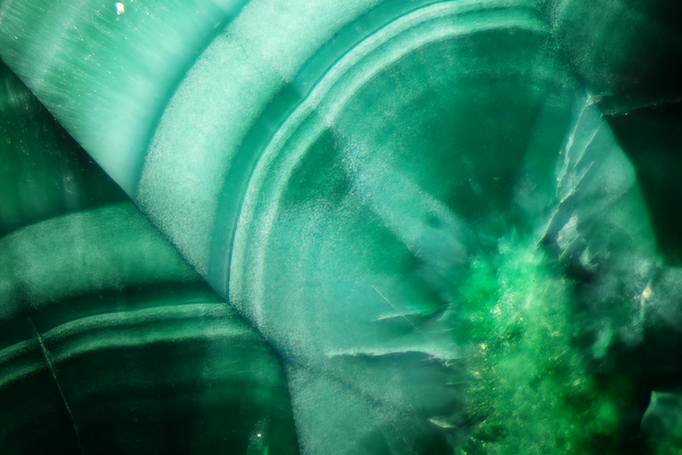
 Figure 6
HPT treated Blue sapphire, crop circles or spiral formations, fiber optic light, FOV 2.0 mm: Photomicrograph by Yenruedee Lhongsomboon
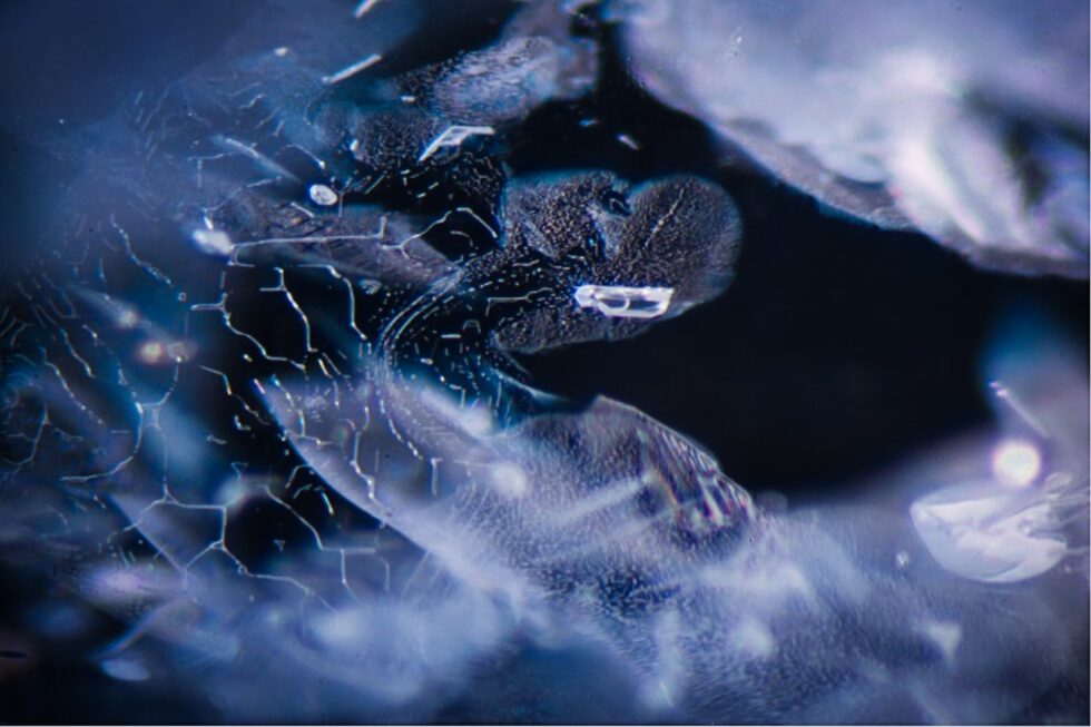
 Figure 7
Be diffusion treated blue sapphire, displays clouds and coiled spring-like inclusions, fiber optic light, FOV 3.6 mm: Photomicrograph by Sarocha Luetrakulprawat
About the writer
Sarocha Luetrakulprawat
Sarocha Luetrakulprawat Sarocha Luetrakulprawat was an Analyst at
ICA IGemLab. She graduated from Burapha University in Chanthaburi, Thailand in Gems and Jewellery at Faculty of Gems. Her responsibility was the identification on gemstones and the operation advanced analytical instrumentation to obtain and analyse the data.